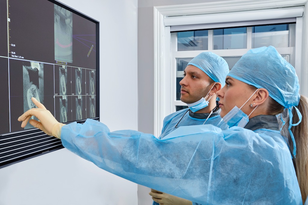
At Gartside Street Dental Lounge in Manchester city centre, our clinicians share practical guidance to help you make confident treatment decisions.
Worry about X-ray radiation is sensible. Radiation has a deserved reputation for being something to take seriously, and modern patients are generally more health-aware than ever. The reassuring news is that dental X-rays sit at the very lowest end of medical imaging in terms of dose — by some distance — and the regulatory framework that governs them in the UK is among the strictest in the world. The aim of this post is to give you the actual numbers, in context, so you can make an informed decision rather than relying on intuition.
A dental X-ray works by passing a brief, tightly collimated beam of radiation through the area of interest onto a digital sensor on the other side. The image appears on screen within a second or two. The exposure time for a single bitewing image is typically around 0.1 to 0.4 seconds. The beam is shaped to cover only the teeth being imaged, and modern sensors are sensitive enough to require very small doses to produce a clear picture.
The doses themselves are best understood in comparison with everyday background radiation. Average UK natural background radiation is roughly 2.7 millisieverts (mSv) per year, mostly from radon gas, cosmic rays, and food. A single dental bitewing delivers around 0.005 mSv. A single periapical X-ray (a longer image of one tooth and its root) is similar. A panoramic OPG, which captures both jaws in one image, delivers roughly 0.01 to 0.02 mSv. A cone-beam CT scan, used for implant planning, delivers around 0.05 to 0.1 mSv. By comparison, a transatlantic flight delivers around 0.08 mSv, and a medical chest CT delivers around 7 mSv.
Translating those numbers into something intuitive: a single dental bitewing is roughly equivalent to one day of natural background radiation, or about one-sixteenth of a single transatlantic flight. A full mouth series of bitewings, taken every couple of years, is the equivalent of perhaps a week of natural background. Even a 3D cone-beam scan for implant planning is on the order of one to two weeks of background radiation. These doses are several orders of magnitude smaller than the levels at which radiation exposure has any measurable health effect.
Digital sensors changed the dental radiation landscape fundamentally. Old-fashioned dental film required roughly ten times the dose of a modern digital sensor to produce a usable image, so figures from older textbooks substantially overstate what patients receive today. Almost every UK private practice, including ours, uses digital sensors. If you have not had a dental X-ray for ten or fifteen years, the dose you receive today is significantly lower than the dose you remember.
Every X-ray taken in a UK dental practice is governed by the Ionising Radiation (Medical Exposure) Regulations 2017, often known as IRMER. The principle behind IRMER is summarised by the acronym ALARA: as low as reasonably achievable. Every X-ray must be clinically justified by the dentist, optimised so that no more dose is used than necessary, and recorded. We do not take routine X-rays "just because" — every image must answer a specific clinical question and the rationale is documented in your notes.
How often X-rays are actually needed depends on your individual risk. Guidance from the Faculty of General Dental Practice and SDCEP is risk-based rather than fixed: bitewings every twelve to twenty-four months for low-risk adults, every six to twelve months for higher-risk adults with active decay or active gum disease. Periapical images are taken only when investigating a specific tooth — pain, suspected abscess, root fracture, or before a planned procedure on that tooth. Panoramic and 3D images are reserved for specific planning needs. If you are stable and low-risk, you may go several check-ups without any X-rays at all.
A reasonable question patients ask is why X-rays are taken at all if the visual examination looks fine. The answer is that around a third of dental decay starts on the contact surfaces between teeth, where it is invisible to the naked eye until it has spread well into the tooth. Bitewing X-rays catch early decay between teeth while it is still small enough to treat with a tiny filling, rather than waiting until it reaches the nerve and becomes a root canal or an extraction. The radiation dose used to detect a small cavity is many orders of magnitude smaller than the consequence of leaving that cavity undiagnosed.
Lead aprons and thyroid collars are standard practice in the UK. Modern collimated beams are so tightly aimed that scatter to the rest of the body is minimal, and current European guidance has actually moved away from routine use of lead aprons in some contexts because they no longer meaningfully reduce dose to most adults. Thyroid collars remain routine because the thyroid gland is more radiosensitive, and many practices, including ours, continue to offer aprons as added reassurance, particularly for younger patients.
Pregnancy is the question we are asked about most often. As a precaution, non-essential X-rays are deferred during pregnancy, and we will always ask if you are or might be pregnant before any imaging. In a true dental emergency — a severe infection, for example, where we need to plan urgent treatment — X-rays can be safely taken with abdominal shielding because the beam path is far from the abdomen and the dose is so small. Routine check-up X-rays simply wait until after delivery.
Children receive even fewer X-rays than adults, and at lower exposure settings. Their developing tissues are slightly more radiosensitive, so doses are reduced and intervals are longer when caries risk is low. Dental X-rays in children are still common and important — early decay in baby and adult teeth is much harder to see clinically — but the principle of justification and minimisation applies even more strictly.
Cone-beam CT, the 3D scan we use for implant planning, deserves a specific mention because the dose is higher than a 2D image. A typical small-field CBCT for a single implant site delivers around 0.05 mSv, less than a long-haul flight. A larger CBCT covering both jaws delivers up to around 0.1 mSv. Even at the higher end this is a fraction of a single medical CT scan. CBCT is taken once per planning episode and provides information that simply cannot be obtained from 2D images, including precise distances to the inferior dental nerve, sinus floor, and adjacent roots.
The cumulative dose from routine adult dentistry is reassuringly small. An adult having two bitewings per year for fifty years would accumulate roughly 0.5 mSv from dental imaging — about two months of natural background radiation, spread across half a century. The cumulative figure simply does not reach the levels at which radiation epidemiology shows measurable risk.
The risk that does deserve attention is the risk of skipping necessary X-rays. Decay between teeth that is missed and untreated will spread to the nerve, leading to abscess, root canal, or extraction. The radiation involved in detecting it early is many orders of magnitude smaller than the harm of leaving it undetected. The conversation worth having with any clinician is not "should you ever take X-rays" but "is this specific X-ray justified for me right now". A good clinician will answer that calmly.
If you have specific concerns — recent medical imaging, pregnancy, a particular medical condition, or simply a preference to discuss before agreeing — please tell us at booking. We use digital sensors, follow IRMER 2017 standards, and are happy to explain the rationale for any X-ray we recommend. You can book a consultation and discuss imaging on the day before any X-ray is taken. Ready to Transform Your Smile?
Take the first step towards your dream smile. Book a free consultation with our expert team at our Manchester clinic and discover how we can help you achieve the confidence you deserve.